Are you suffering from a potential Lisfranc injury?
The Omaha Foot & Ankle Specialists at MD West ONE are able to properly diagnose and treat Lisfranc (midfoot) injuries through both surgical and non-surgical treatments. If you have the following symptoms, you may want to make an appointment with one of our Board Certified Specialists.
- Pain around the region of the arch of the foot
- Swelling on the top of the foot
- Bruising on the top and bottom of the foot. Bruising on the bottom of the foot in the region of the arch is highly suggestive of a Lisfranc injury
- Pain that worsens with standing, walking, or attempting to push off the affected foot. The pain can be so severe that crutches may be required.
Meet MD West ONE's foot and ankle specialists and learn more about how they treat a Lisfranc injury.

Kathleen A. Grier, M.D.
Foot & Ankle Specialist

Scott T. McMullen, M.D.
Foot & Ankle Specialist

David J. Inda, M.D.
Foot & Ankle Specialist

Shane Schutt, M.D.
Foot & Ankle Specialist
Lisfranc Injury Causes, Treatments & Surgery
What are Lisfranc Injuries?
Lisfranc injuries are the result of bones in the midfoot that is broken or ligaments that support the midfoot that is torn. The severity of the injury can vary significantly from tearing a single ligament too complex, involved in many joints and bones in the midfoot. A Lisfranc injury is often misdiagnosed as a simple sprain, especially if the injury is a result of a straightforward injury such as a twist or fall. However, a Lisfranc injury is very different than a simple sprain and should not simply be walked off. It is an injury that often requires surgery and may take many months to heal.
The midfoot joints (Lisfranc joints) can be injured with both low and high energy injuries. Low energy injuries can happen with a simple twist or fall. This is commonly seen in football and soccer players, but can also happen if someone stumbles over the top of a flex foot during everyday activities. More severe injuries often occur from direct trauma, such as a fall from height or a car accident. These high-energy injuries can cause multiple fractures and dislocations of the joints. As a result, the cartilage that covers the end of the bones in the joints can be damaged. This can lead to increased stress across the midfoot joints which can result in both collapses of the arch and arthritis.
Anatomy
The bony structure of the midfoot is composed of joints of the wedge-shaped metatarsal bones that extend out toward the toes with their corresponding cuneiform bones. Important features of the boney anatomy include:
- The cuneiforms and metatarsal bones have trapezoidal configurations, with the second metatarsal base in the middle cuneiform serving as the keystone of the transverse arch, much like a Roman arch.
- The middle cuneiform is recessed, allowing the second metatarsal base to lock into the joint in a mortise configuration.
- The individual joints of the midfoot are flat with very little inherent stability making the soft tissues that hold them together extremely important.
The bones in midfoot are held in place by connected tissues called ligaments that stretch not only across the foot but also down the foot. There is no ligament connecting the first metatarsal with the second metatarsal. As a result, a twisting fall or other traumatic injuries such as a car accident can break and shift these bones out of place. The interosseous ligament commonly referred to as the Lisfranc ligament, is the strongest and most important ligamentous restraint of the midfoot.
The midfoot is critical in stabilizing the arch during walking and jumping activities. The midfoot transfers the force is generated by the calf muscles to the front of the foot allowing the heel to rise and propel the body forward.
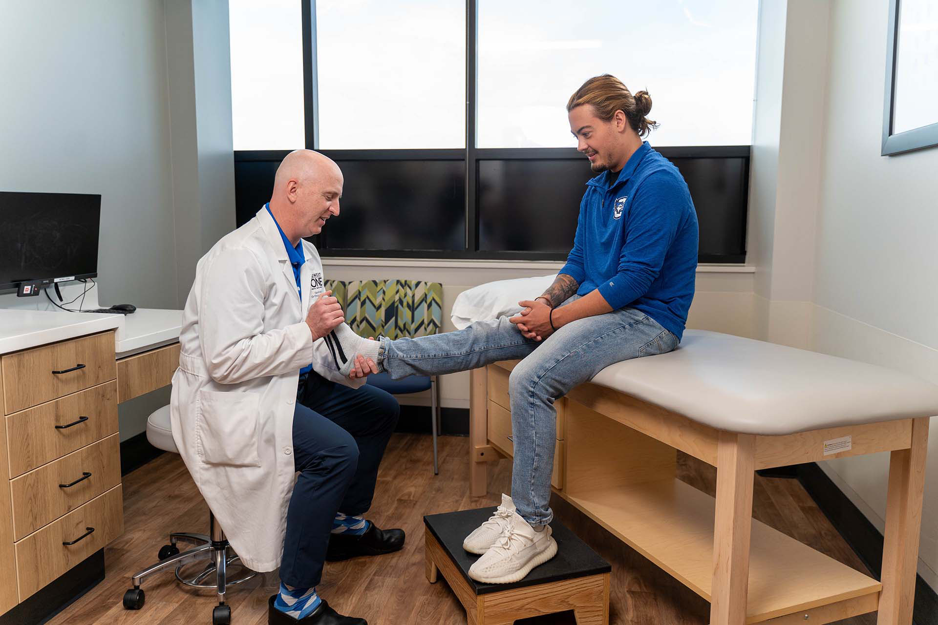
What will be looked for during a physical examination
- Looking for a bruising along the bottom of your foot, suggesting a tear of the Lisfranc ligament or a fracture of the tarsal or metatarsal bones.
- Tenderness to palpation along the top and bottom of the midfoot.
- Pain with stress examination of the midfoot, consisting of gently stabilizing the heel and twisting the front of the foot to evaluate for pain and instability in the midfoot.
- Single leg heel-rise test and single-leg stance. With this, the doctor may ask you to stand on one foot and then come up onto your tiptoes to put stress across the midfoot. If the midfoot is unstable, this test could cause pain compared to the uninjured side.

Tests
- X-Rays: Fractures of the metatarsal’s and tarsal bones can be evaluated using non-weight-bearing x-rays. Moreover the position of the joints can often be evaluated. Any change in the normal joint position may suggest an injury to the joint capsules and ligaments that hold them in place.
- Weight-Bearing X-Rays: If the injury happened from a relatively low energy mechanism, such as from a simple twist or a fall, x-rays may be taken with the patient standing. In this instance, the doctor is looking for damage to the ligaments that stabilize the midfoot. While the ligaments themselves cannot be seen on x-ray, ligament tears can result in instability or shifting of the joints.
- MRI Scan: The doctor will order an MRI scan in order to better visualize the soft tissue including the tendons and ligaments. This is often done in situations where the diagnosis is not readily apparent on x-rays, but the patient is unable to bear weight on the foot.
- CT Scan: CT scan is a more detailed x-ray that can get a cross-sectional image of the foot. Often times physicians use this for surgical planning as it provides a better picture of the injury and the number of joints that have been involved.
Treatment
Treatment of a Lisfranc injury depends on the severity of the injury and whether it has resulted in any instability in the midfoot. Unstable injuries often require surgery, while stable injuries can be treated without surgery.
Non-Surgical Treatment Options
When there are no fractures or dislocations of the joints, nonsurgical treatment may be that all that is necessary. In this situation the ligaments have not been completely torn, and heal on their own as long as they are protected. I nonsurgical treatment plan often includes wearing a non-weight-bearing cast or boot for six weeks. Weightbearing is then slowly progressed in a boot or a cast over the next 4 to 6 weeks until the patient has full weight-bearing around three months after the injury. Occasionally, recovery will also include using orthotic arch support as the patient progresses toward normal activities. During the recovery process, the doctor will arrange regular follow-ups and take additional x-rays to make sure the foot is healing well. If there is any evidence that the bones have shifted or moved during the recovery period, then surgery will be needed to realign and stabilize the bones.
Surgical Treatment Options
Surgical treatment is recommended for all injuries with a fracture that extended the joints of the midfoot or with any malalignment of the joints. The goal of surgical treatment is to realign and stabilize the joints and return the fractured bone fragments to a normal alignment.
Open reduction, internal fixation: In this procedure, the bones of the midfoot are positioned correctly (reduced) and held in place with plates and screws allowing them to heal in the appropriate position. During this surgery, plates and screws are placed across the joints that have some degree of motion. For this reason, some or all of the hardware may be removed later as it can weaken and break over time. If the surgeon plans to remove the hardware, it will often be done be between three and five months after the initial surgery. Occasionally, the hardware can be left in place if it does not bother the patient.
Fusion: Certain types of Lisfranc injuries that damage only the ligaments, or result in a significant amount of cartilage damage, a fusion may be recommended as the initial surgical procedure. In a fusion, the bones grow together so that they heal into a single, solid piece.
The joints of the midfoot often involved in a Lisfranc injury have very little motion and mainly transfer stress from the front of the foot to the hindfoot and ankle. Fusing the joints, therefore, can result in a very normal gait. Because the bones grow together to single, solid piece, the plates and screws will not weaken and do not need to be removed.
Recovery
After either an open reduction, internal fixation surgery, or fusion surgery, a period of non-weightbearing for 6 to 8 weeks is recommended and either a cast or a boot. After the initial weight-bearing portion, the doctor will allow slow, progressive weight-bearing initially in a boot and subsequently in a supportive tennis shoe. If the patient had an open reduction, internal taxation surgery, impact activities including running and jumping should be avoided until the hardware has been removed.
Most recreational athletes are able to return back to their pre-injury level. However, these injuries can often damage the joint cartilage which may ultimately result in arthritis, pain, and progressive deformity what time. In these situations, an additional surgery to fuse the midfoot may be required.

American Orthopaedic Foot & Ankle Society
All of the foot and ankle surgeons in the practice are recognized members of the American Orthopaedic Foot & Ankle Society. It is the oldest and most prestigious medical society dedicated to the foot and ankle. The mission of the society is to advance science and practice of foot and ankle surgery through education, research, and advocacy on behalf of patients and practitioners. These physicians dedicate their time and energy to improving the patient experience and their knowledge in their field. For more information visit http://www.aofas.org.
MD West ONE Foot & Ankle Specialists:
The Foot & Ankle Specialists are all Board Certified and Fellowship-Trained, meaning they’ve focused their education, training and research on orthopaedic surgery of the foot and ankle.
